A Low Cost Teleradiology Workstation
M. Desco, J. López, H.
Rahms, F. del Pozo, P. García-Barreno
Unidad de Medicina Experimental, Hospital General
Universitario "Gregorio Maraёón"
Madrid, Spain
OBJETIVES
To develop a low cost workstation oriented to the
digitization of multi-modality medical images, their storage and
teletransmision, with internal archive and data communication based opn DICOM
3.0, teleconference services, modular structure and possibility of direct
connection with proprietary systems. Opcional services are patient management,
and long-term archive on CDR.
The product must be transferred to the national
industry in order to proceed to its commercialization by the end of 1998.
MATERIAL AND METHODS
Image aquisition
devices: ааSPECT: Siemens Orbiter-75
ааааааааааааааааааа ааааааааа ааааааааа PET:
Elscint Posicam HZL
ааааааааааааааааааа ааааааааа ааааааааа MRI:
Philips Gyroscan 0.5T y 1.5T
ааааааааааааааааааа ааааааааа ааааааааа CT:
Philips LX y Siemens Somatom.
Programming tools:а IDL 5.0
(Windows y Mac) environment, with some routines in C language.
RESULTS
An application as been implemented according to
the following specifications: Image capture: Scanner (8-12 bits) and video
frame grabber (up to 1024 x 1024). Compatibility: Allowable image formats:
Interfile, DICOM 3.0, and some proprietary formats (PHILIPS, PET Posicam, GE
Signa). Storage: SQL database and DICOM; CDR recording for final archiving. Telecommunication:
Remote database access and on-line/off-line image transmision; cooperative
work, videoconference. Visualization: 2D presentation including configurable
screen layout, Zoom/Pan, Level and window, user defined color LUTs. Quantification
tools. 3D volume navigation though orthogpnal planes, 3D rendering. Medium and
high resolution monitors allowable. Reporting: Both voice and conventional
reports can be attached to the patient studies.
User Interface has been specially designed to be
as simple possible, and validated with clinical users.
SUINSA S.A., a national medical equipment
manufacturer, faces the commercialization of the system in a short term.
 |
 |
|
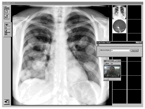
Figure 1: Program layout, showing a thorax study and a videophone.
|
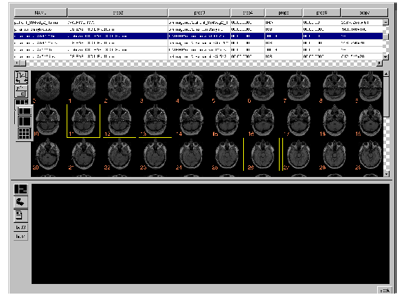
Figure 2: Image selection tool.
|
There are few commercially available platforms
offering the same wide range of features. The application presented here is
portable, simple, low cost and modular design, allowing for specific
customizations according to the particular user requirements.
Corresponding
author:
Sr. Manuelа
Desco
Unidad de Medicina Experimental.
Hospital General Universitario "Gregorio
Maraёón"
Madrid Spain
e-mail: desco(at)mce.hggm.es
Oral presentation at EuroPACS'98, Barcelona, Spain