A Low Cost Multimodality Workstation
M. Desco, J. López, J. Lampreave, C. Benito, A. Santos, P. García-Barreno
Unidad de Medicina Experimental. Hospital General Universitario "Gregorio Marańón"
Madrid, Spain
OBJECTIVE
To develop a workstation for processing and visualization of medical multimodality image information. The program must be able to import images from different sources (MRI, SPECT, PET, CT), providing the necessary tools for the registration, fusion. quantification and visualization of results, pursuing the final goal of making it easier the joined interpretation of morphologic and functional studies.
The platform must necessarily be low cost, and based on standards as much as possible.
IMPLEMENTATION
Modalities and acquisition devices: SPECT: Siemens Orbiter-75; PET: Elscint Posicam HZL; MRI: Philips Gyroscan and General Electric Signa; CT: Philips LX y Siemens Somatom. DICOM media files import.
Development tools: IDL 5.0 programming environment (Windows y Mac) and 'C' language.
RESULTS
A software product has been designed and implemented with the following characteristics: Communications: Image formats: Interfile, DICOM, and proprietary formats (PHILIPS, PET Posicam, GE Signa). Visualization: 2D and 3D presentation according to orthogonal planes, Zoom/Pan, Level and Window, different color LUTs. Image processing: Segmentation: Manual, threshold, region growing, clustering. Image quantification. 3D Registration Intrasubject/Intersubject; Landmark based and totally automated (AIR) algorithms. Image fusion: by semitransparent coloured overlay.
This application is under clinical validation in different research projects. Up to now, it has been used with more than 100 patients (SPECT, PET y MRI) with different pathologies, mainly epilepsia and schizophrenia.
DISCUSSION AND CONCLUSIONS
The few commerial tools offering similar features are expensive and difficult to integrate in multi-manufacturer environments. Our system is demonstrating excellent results as a tool for the joined interpretation of anatomical and functional studies, offering a very good connectivity at a low cost.
 |
||
|
Figure
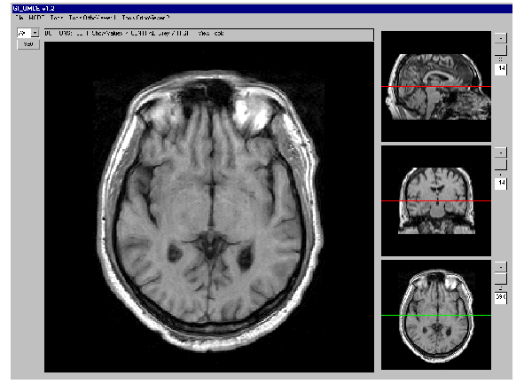
1: Program layout
|
Figure
2: Segmentation tool
|
Figure
3: Fusion tool
|
Corresponding author:
Sr. Manuel DescoOral presentation at EuroPACS'98, Barcelona, Spain