Athanasios M. Demiris,
Carlos E. Cárdenas S., H.P. Meinzer
Deutsches
Krebsforschungszentrum - Div. Medical & Biological Informatics -
Heidelberg, GERMANY
Objective
We want to denote the importance of componentware for medical imaging and introduce an architecture that establishes the necessary infrastructure for the creation of domain-specific software components.
Introduction
In the past years there has been a clear demand for added-value functionality in viewing stations. Added-value functionality can help deal with specialized medical problems such as volumetric calculations in CT-images of the upper abdomen, regurgitant jets measurements in US-images of the heart etc. One approach is to install at the end-user site, i.e. the radiological department, a monolithic image processing system dealing with all tasks in a function-oriented way and trying to a-priori cover all possible cases by integrating a maximum of image processing and visualization functions. The major disadvantage of such systems is that they are closer to the image processing and farther from the medical way of thinking thus introducing a higher work-load for the medical end-user. The alternative approach is the creation of many specialized, task-oriented software modules dealing with each new clinical problem separately. This sometimes leads to functional overlap between modules, e.g. a region growing algorithm would be integrated in more than one modules, but enforces user-centered design and developments as well as encapsulation of overcomplicated functionality and paramerterization which is feasible only when a concrete and restricted application domain is addressed.
While the specialized modules vary from case to case, it is obvious that there are some tasks that remain invariant, e.g. the image handling and communication. This invariant functionality can be provided by a host system, e.g. a viewing station or a tele-radiology system, that is customizable and can be extended by integrating specialized components. In this work we will present an information architecture delivering the infrastructure needed for the easy creation of specialized components for viewing stations or tele-radiology systems. We identified the development phases for medical imaging components in terms of developer’s activities, we introduced inter-operating supporting facilities for each activity, designed the structured storage and retrieval along with meta-information about such components and implemented a first version of this architecture by means of object-oriented technology.
System Description
The scenario which led to the identification of the development activities was observed and analyzed within our imaging group and the clinical environment of the Heidelberg-University-Clinics. According to this scenario medical end-users contact software developers of our group and ask for digital support for a new clinical problem. The developer would then
1. first become acquainted with the new data, then
2. browse the algorithms-arsenal in order to find the most appropriate tools, subsequently
3. estimate the parameter sets that would allow the selected algorithmic material to operate successfully on the new data, after that
4. create a graphical user interface for accessing the selected functionality and finally
5. either integrate the developed solution into an existing system or ship a stand-alone version into the clinical environment.
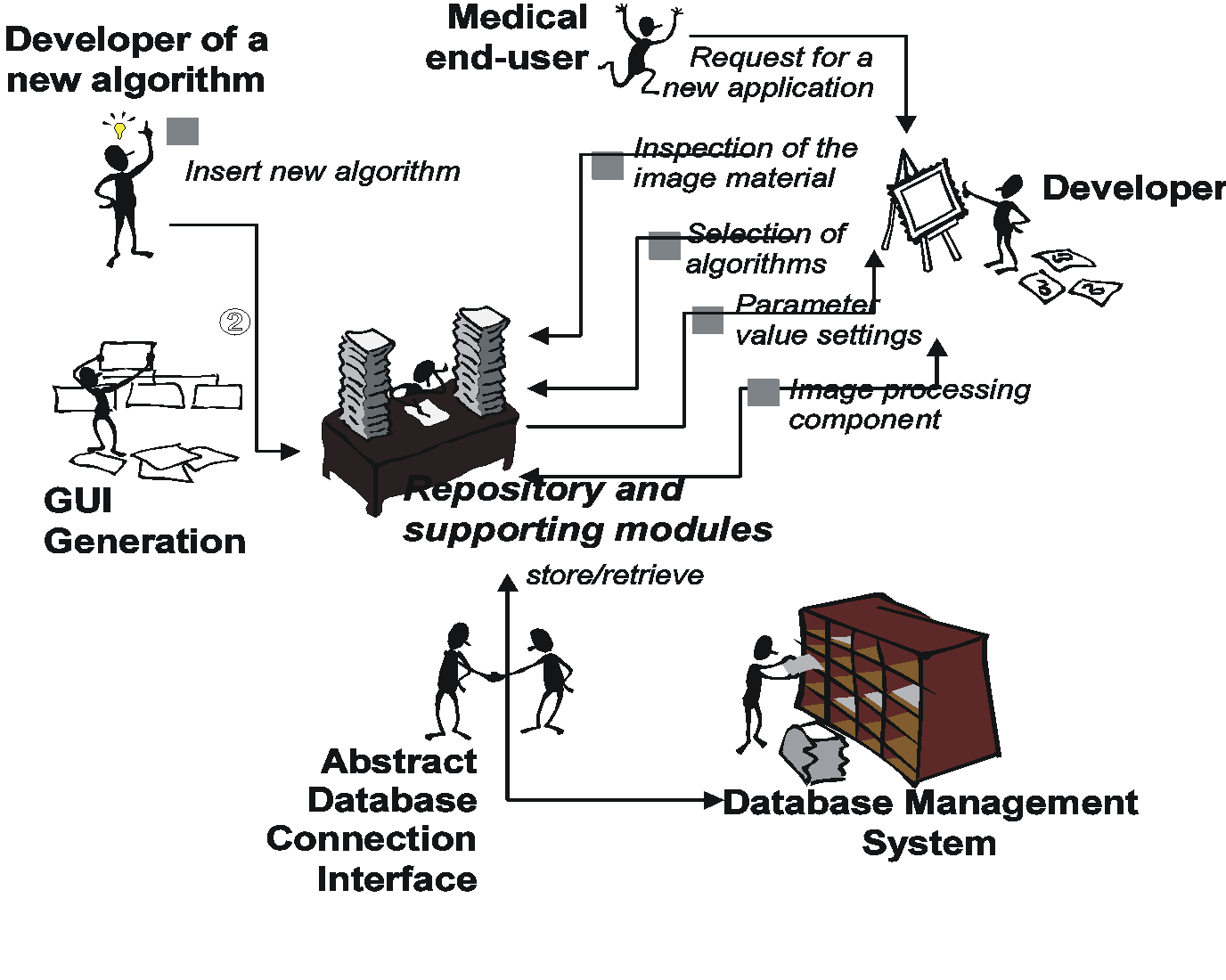
Our architecture contains as its central part a repository for imaging algorithms. The repository enables a structured storage of imaging algorithms along with meta information about their functional aspects, side effects, administrative and execution data etc. and supports an intuitive, goal-oriented query. ”Around” the repository there are three more modules in the architecture designated to deal with the other development aspects:
· one module helps the developer obtain statistical values for the new image material by analyzing selected characteristics of given samples,
· a second component contains a fuzzy-logic based rule engine that helps estimate optimal parameter values for a given algorithm, based on statistical properties of the images and the parameter constraints of the selected algorithm itself, and finally
· a module that automatically creates graphical user interface elements for the manipulation of the algorithms.
Results
The overview of the architecture can be seen in figure 1. We implemented the architecture using object-oriented componentware techniques (general concepts, Java-Bean technology etc.). The modules of the architecture are implemented as object-oriented frameworks, thus being fully customizable and extensible. They can be used as domain-specific extensions to other existing development support environments (CASE) and not as a replacement of the latter. The repository can accommodate algorithms written in several programming languages, e.g. C, C++, Java, Smalltalk, and virtually all platforms. The openness of the architecture is guaranteed by the use of standards during development, i.e. the CDIF/MOF-like structure of the repository.
Figure 1: The overview of the architecture: creation of a new system based on existing components in the repository and support by the other modules (steps 1,2, 3, gamma) and insertion of new algorithmic material in the repository with the automatic creation of graphical elements for later retrieval (steps 1,2).
Corresponding Author:
Athanasios M. Demiris
Deutsches Krebsforschungszentrum
Div. H0100 Medical & Biological Informatics
Im Neuenheimer Feld 280 D-69120 Heidelberg
Phone: +49-6221-422328 FAX: +49-6221-422345
E-Mail: A.M.Demiris(at)dkfz-heidelberg.de
Oral presentation at EuroPACS'98, Barcelona, Spain