A MINIPACS DESIGN FOR THE VIEWING OF DIGITAL MAMMOGRAPHIES
P.G. Rodriguez1,2, L.Gomez2, R.G.Carrión2, E.Cernadas1, A.Plaza1 and J.J.Vidal3
1 Dpto. Informática, Escuela Politécnica, University of Extremadura, Spain, 2 Lab. Imaxe Dixital, Dpto. Electrónica e Computación, University of Santiago de Compostela, 3 Lab. de Radioloxía, Hospital Xeral de Galicia, University of Santiago, Santiago de Compostela Spain.
Abstract:
Nowadays many efforts are being focused on design of general systems to manage medical images. Conventional mammography is currently the most efficient technique to detect early breast cancer. Due to special requirements of breast radiography images, integration of digital mammography in PACS is not yet achieved. In this paper, we present a possible solution with a specific miniPACS design to mammography.
PACS is currently changing the way of working in clinical environments1. But the whole integration in hospitals of these digital technologies could be reached by using specific miniPACS2. Owed to peculiar characteristics of mammographic images, miniPACS can be adequate to solve archiving, transmission, viewing and other limitations of conventional mammography in relation with detecting signs of early breast cancer (masses and microcalcifications)3. Thus, new topics like telemammography4 and CAD (Computer-Aided Diagnosis)5,6 would be easily introduced to radiologists. Nevertheless, their development is strongly related to hardware technology advances. In any case, we believe that preservation of optical image quality is the most important aspect to take into account in a mammographic miniPACS design in order to reach acceptation among radiologists.
MATERIALS AND METHODS
A friendly system design needs to combine computer sciences and psychology, focusing on user perception. The main point to take into account in this case would be the radiologist-computer interaction7,8. So, several systems with different philosophies have been proposed to be used in clinic environments.
Seven years ago, a text mode user interface has been developed with a pull-down menu on a PC monitor and a bit-slice processor: first using a low resolution (512x512 pixels) and secondly with a high resolution (1024x1024) TV monitor to display mammographies.
A few years later, a second system over a RISC workstation with a high resolution 20” monitor for displaying a GUI (Graphical User Interface) and mammographies was implemented, which leads to a higher interaction between workstation and radiologists.
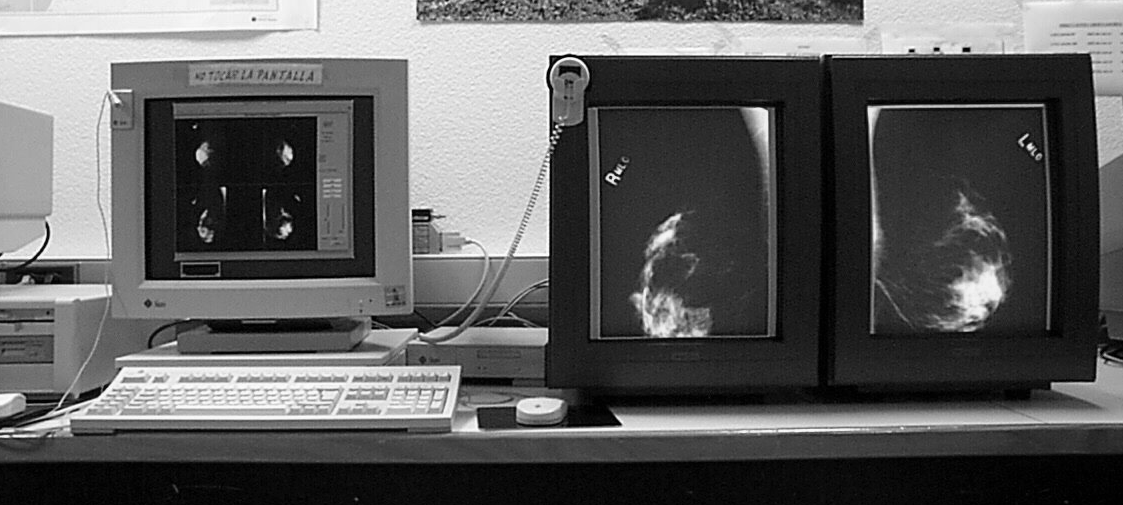
The current system consists of two specific black and white MegaSCAN 2Kx2.5Kx10bits monitors with DOME controllers, working over a SunSPARC20 workstation under the Solaris operating system. A friendly user interface and hierarchical mammographic database with three levels (patients, studies and views) have also been developed under OSF/Motif (figure). Several mammographic viewing tools have been implemented, such as window/level controls, zoom and roam, ROIs selection, etc. The first three functions have been implemented using the three-button mouse. So, interaction is improved, since radiologists do not need to look away from the screen to manage the above mentioned tools. Furthermore, both a local non-deforming automatic enhancement technique based on Unsharp Masking filters9, and a specific lossless wavelet algorithm for image compression10 have been developed.
A continuous evaluation of systems is needed in the introduction of new technologies in hospitals. Statistical and practical studies over the LLNL mammographic image database with microcalcifications from UCSF11 have been done in order to test our system.
Results and discussion: evolution
In order to preserve radiography quality, high spatial and gray level resolution should be used in the digitalization process. This fact normally involves that powerful machines and special devices are required in archiving, transmission and viewing processes.
The first system had computational power, portability and image quality limitations. RISC architecture of the second system increases computational power, while the use of UNIX and OSF/Motif improves its portability. In addition, the GUI leads to a higher radiologists-computer interaction. However, image quality is not enough for radiologists, so the current system is trying to solve it, at the expense of using a high-cost specific hardware again.
Archiving and transmission are optimized by our specific compression module. Viewing is improved by the specific mammographic tools and the use of high resolution monitors. At the moment, all the utilities are only possible if developing specific designs (miniPACS) with a intuitive GUI.
Nevertheless, one critical issue in transmission mammographic images continues to be communication protocols among different devices in hospitals. Nowadays, the DICOM (Digital Imaging and Communication in Medicine, or ACR/NEMA 3.0)12 standard for medical images is under development. But DICOM is not yet standardized for mammographic viewing, due to its special image high quality requirements.
To summarize, philosophies and tendencies in system designs are changing across time. On the one hand, portability is desirable, which frequently implies the use of general hardware systems. On the other hand, image quality is necessary, but this aim is only reachable at the expense of high hardware cost or general hardware technology advances. Its future installation in hospital will need more screens and speed to display mammographies in diagnosis.
CONCLUSION
Our experience has shown that MiniPACS could be an approximation to introduce new advances in clinical environments. Radiologists should be enticed to use these kinds of systems. However, mammographic image quality needs to be improved by technology in order to achieve acceptation among radiologists.
REFERENCES
[1] Osteaux M.: "A Second Generation PACS Concept". Ed. Springer-Verlag, 1992.
[2] Horii S., Feingold E., Coleman B., Arger P., Langer J., Jacobs J., Nisenbaum H., Rowling S., Seshadri S.: "The Use of MiniPACS Technology in Ultrasound: the Potential for Productivity Improvement", PACS Design and Evaluation, Proc.SPIE, Vol.2435, 257-262, 1995.
[3] Säbel M. and Aichinger H.: “Recent developments in breast imaging”. Phy.Med.Biol.41, 315-368 (1996)
[4] Goldberg M.A., Dwyer III S.J.: "Telemammography: Implementation Issues", Telemedicine Journal, Vol.1, No.3, pp.215-226, 1995.
[5] Jiang Y., Nishikawa R.M., Metz C.E., Wolverton D.E., Schmidt R.A., Papaioannou J., Doi K.: "A computer-aided diagnostic scheme for classification of malignant and benign clustered microcalcifications in mammograms", Digital Mammography’96, Ed. Elsevier Science, 1996.
[6] Méndez A.J., Tahoces P.G., Lado M.J., Souto M. and J.J.Vidal: "Computer-aided diagonosis: detection of masses on digital mammograms", Proceedings IWISO´96, Manchester, UK, Nov.1996. Ed. Elsevier.
[7] Astley S. et al.: “Automation in mammography: computer vision and human perception”. International Journal of Pattern Recognition and Artificial Intelligence, Vol.7, No.6 (1993) 1313-1338.
[8] Thimbleby H.: "User Interface Design", Addison-Wesley Publishing Company, 1990.
[9] Cernadas E., Gómez L., Rodríguez P.G., Casas A., Carrión R.G. and Vidal J.J.: "Design of unsharp masking filters in the frequency domain: Parametrization for breast radiographs" pp.463-466 in Digital Mammography'96. Excerpta Medica. International Congress Series 1119. 1996 Elsevier Science B.V.
[10] Carrión R.G., Rodríguez P.G., Gómez L., Cernadas E., and Vidal J.J.: “Compression Techniques Adapted to Digital Mammography: A comparative study”, Radiological Society of North America 1997, Chicago, USA, December 1-5, 1997, pgs. 523.
[11] Mascio L.N., Frankel S.D., Hernandez J.M., Logan C.M.: "Building the LLNL/UCSF Digital Mammogram Library with Image Groundtruth", 3r International Workshop on Digital Mammography, Chicago, p.57, June 1996.
[12] Bidgood W.D. and Horii, S.C.: "Introduction to the ACR-Nema DICOM Standard", Radiographics, 12 (3), pp.537-548, 1992.
Corresponding author:
Pabro García Rodriguez
Dpto. Informática, Escuela Politécnica,
University of Extremadura,
10071 Cáceres (Spain).
E-mail: pablogr(at)unex.es
Oral presentation at EuroPACS'98, Barcelona, Spain